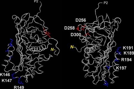
FIGURE 7.
Three-dimensional structure of human PEDF (from Protein Data Bank ID 1IMV) to illustrate the location of the HA-binding site. The two structures are rotated about 180° from each other with highlighted positions of single alterations made in this study. In blue are basic amino acids Lys146, Lys147, and Arg149 residues located in a turn between β-strand s2A and α-helix E, and Lys189, Lys191, Lys194, and Arg197 located in another turn between α-helix F and β-strand s3A, both within BX7B HA-binding sites; in red are acidic amino acids Asp256, Asp258, and Asp300 corresponding to the collagen-binding site. P2 corresponds to the residue next to the homologous serpin reactive site, P1; and N– in yellow indicates the position of the amino-end terminus of the polypeptide in the three-dimensional structure corresponding to position 26. Structures were visualized and reproduced using Cn3D (NCBI).

