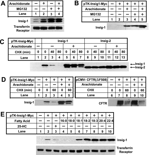
FIGURE 1.
Unsaturated fatty acids inhibit degradation of Insig-1. A, CHO-7 cells were seeded at 3.5 × 10⁵/60-mm dish on day 0. On day 2, cells were switched to medium A supplemented with 5% delipidated FCS with 50 μm compactin and 50 μm mevalonate. On day 3, 100 μm arachidonate or 10 μm MG132 was added into the medium as indicated. After incubation for 6 h, cells were harvested. Cell lysate was subjected to SDS-PAGE followed by immunoblot analysis with anti-Insig and anti-transferrin receptor. B, SRD-13A cells were seeded as described in A. On day 2, cells were transfected with 0.4 μg of pTK-Insig1-myc. Total plasmid concentration was adjusted to 2 μg/dish by using the empty vector pcDNA3.1. Following incubation for 8 h, cells were switched to medium A supplemented with 5% delipidated FCS. On day 3, cells were treated as described in A, and transfected Insig-1 was detected by immunoblot analysis with IgG-9E10. C, SRD-13A cells were seeded, transfected with 0.4 μg of pTK-Insig1-myc or pTK-Insig2-myc, and incubated as described in B for the first 2 days. On day 3, cells were incubated in the absence or presence of 100 μm arachidonate in medium supplemented with 5% delipidated FCS for 6 h. These cells were then treated with 50 μm cycloheximide (CHX) for the indicated period of time. Cell lysate was subjected to SDS-PAGE followed by immunoblot analysis with IgG-9E10 to detect transfected Insig proteins. D, SRD-13A cells were seeded, transfected with 0.4 μg of pTK-Insig1-myc or 1.5 μg of pCMV-CFTR(ΔF508), and treated with arachidonate and cycloheximide (CHX) as described in C. Cell lysate was subjected to SDS-PAGE followed by immunoblot analysis with IgG-9E10 (against Insig-1) and anti-CFTR. E, SRD-13A cells were seeded, transfected with 0.4 μg of pTK-Insig1-myc, and incubated as described in B for the first 2 days. On day 3, cells were treated with a 50 μm concentration of the indicated fatty acids or 1 μg/ml 25-hydroxycholesterol (25-HC) in medium supplemented with 5% delipidated FCS for 6 h. Cells were then harvested, and cell lysate was subjected to SDS-PAGE followed by immunoblot analysis with IgG-9E10 (against Insig-1) and anti-transferrin receptor.

