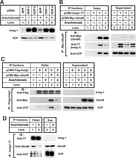
FIGURE 5.
Ubxd8 mediates the complex formation between VCP and Insig-1. A, HEK-293S/pInsig1 cells were seeded, transfected with indicated siRNA, and treated as described in Fig. 4B. Cell lysate was subjected to SDS-PAGE followed by immunoblot analysis with anti-VCP and anti-T7 (against Insig-1). B, SRD-13A cells were seeded, transfected, and treated with arachidonate as described in Fig. 3C. Detergent lysates of these cells were subjected to immunoprecipitation with anti-Myc IgG-coupled agarose beads to immunoprecipitate transfected Ubxd8. Pellets (representing a 0.1 dish of cells) and supernatants (representing a 0.01 dish of cells) of the immunoprecipitates were subjected to SDS-PAGE followed by immunoblot (IB) analysis with polyclonal anti-Myc (against Ubxd8), anti-T7 (against Insig-1), and anti-VCP. C, SRD-13A cells were seeded, transfected with 0.2 μg of the indicated plasmids, and treated with arachidonate as described in Fig. 3C. Detergent lysates of these cells were subjected to immunoprecipitation (IP) with anti-FLAG IgG-coupled agarose beads to immunoprecipitate transfected Insig-1. Pellets and supernatants of the immunoprecipitates were analyzed as described in B. D, HEK-293S/pInsig1 cells were seeded at 6.0 × 10⁵/100-mm dish on day 0. On day 2, cells were switched to medium containing 10% delipidated FCS. On day 3, 10 μm MG132 was added into medium in the absence or presence of 100 μm arachidonate. After incubation for 6 h, cells were harvested, and the cellular lysate was subjected to immunoprecipitation with anti-T7 to precipitate Insig-1. Pellets (representing one dish of cells) and supernatants (representing a 0.067 dish of cells) of the immunoprecipitates were subjected to SDS-PAGE followed by immunoblot analysis with anti-VCP, anti-Ubxd8, and anti-T7 (against Insig-1). GFP, green fluorescent protein.

