FIGURE 5.
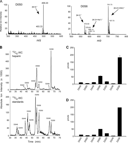
GRIL-LC/MS quantitation of porcine intestinal mucosal heparin. Disaccharides derived from heparin were tagged with [12C6]aniline and mixed with [13C6]aniline disaccharide standards (25 pmol each). A, the mass spectra for a representative low abundance disaccharide, D0S0, and a higher abundance disaccharide, D0S6, are shown. Note the 6 atomic mass units difference between the [12C6]aniline-tagged residue and the corresponding [13C6]aniline-tagged standard for the free molecular ions ([M-H]-1), the sodium adducts ([M-2H+Na+]-1), and the ion pairing reagent adducts ([M-2H+DBA]-1). Adduction ions are typically detected for disaccharides with two or more sulfates (compare the mass spectra for D0S0 and D0S6, see Table 1). B, the individual XIC profiles for the [12C6]aniline-tagged sample and [13C6]aniline-tagged standards for one of three separate analyses. The XIC traces were generated using the m/z values for HS disaccharide standards listed in Table 1. C, GRIL-LC/MS determined disaccharide composition of porcine heparin (n = 3), error bars represent the standard error. D, analysis of an identical sample using the anion-exchange high performance liquid chromatography with post column derivatization and fluorescence detection. The preparation of heparin used in this study did not contain measurable amounts of D0H0, D0H6, D2H0, or D2H6 detected by either LC/MS-GRIL or post column derivatization with 2-cyanoacetamide. The rare disaccharide D2A0, which is also detectable by GRIL-LC/MS (Fig. 3A), was not analyzed in this experiment.

