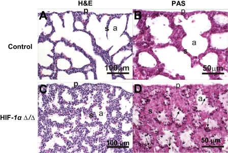
FIGURE 3.
Pulmonary histopathology. Light photomicrographs of hematoxylin and eosin-stained lung sections from control (A) and HIF1αΔ/Δ (C) pups. The lung from HIF1αΔ/Δ pup has less alveolar airspace (a) and thicker alveolar septa (s) compared with control. Large cuboidal epithelial cells line the alveolar septa of HIF1αΔ/Δ pup, whereas the alveolar septa of control pup is lined primarily by squamous epithelial cells (type 1) and only a few widely scattered cuboidal cells (type II). PAS-stained lung sections from control (B) and HIF1αΔ/Δ (D) pups. Cuboidal epithelium lining alveolar septa in HIF1αΔ/Δ pup has greater PAS-stained glycogen (arrows) than that of the control pup (D). All doxycycline treatments were at least 12 days.

