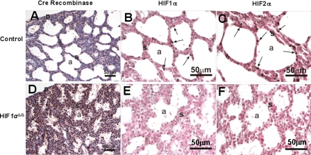
FIGURE 7.
Immunohistochemistry for Cre recombinase, HIF1α, and HIF2α. Lung sections from control (A-C) and HIF1αΔ/Δ pups (D-F) were immunostained for Cre recombinase (A and D), HIF1α (B and E), and HIF2α (C and F). Immunohistochemistry for Cre recombinase shows strong staining of HIF1αΔ/Δ mouse alveolar epithelial cells (D, brown color). Positively stained cells for HIF1α and HIF2α are depicted by solid arrows. HIF1αΔ/Δ pups show decreased expression of HIF1α and HIF2α as compared with control littermates. a, air space; p, pleural surface; s, septa. All doxycycline treatments were for at least 12 days.

