FIGURE 3.
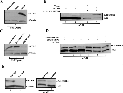
SCCRO augments cullin neddylation in vivo. A, Western blot on HeLa lysates showing elevated SCCRO protein levels in SCCRO transfected (lane 3) relative to untransfected (lane 1) or vector-transfected (lane 2) cells. B, Western blot showing a higher level of neddylated cullins in lysates from SCCRO transfected (lane 2) relative to vector-transfected cells (lane 1). Western blot on HeLa lysates from B after addition of neddylation components (E1, E2, NEDD8, and ATP) showing increased neddylated Cul1 levels in SCCRO-transfected (lane 4) relative to empty vector (lane 3)-transfected cells. C, Western blot on lysates from SCC15 cells showing a decrease in SCCRO protein levels in cells transfected with specific RNAi against SCCRO (lane 3) relative to untransfected (lane 1) or scrambled RNAi-transfected cells (lane 2). D, in vitro neddylation reaction of the same lysates showing decreased Cul1 neddylation in SCCRO-RNAi transfected (lane 3) compared with untransfected (lane 1) or scrambled RNAi (lane 2)-transfected cells. The addition of recombinant SCCRO to the lysate from SCCRO RNAi-transfected SCC15 cells (lane 6) recovers Cul1 neddylation to levels observed in controls (lanes 4 and 5). E, Western blot showing the absence of detectable SCCRO protein in testis lysates from SCCRO–/– mice (left panel, lane 2) in contrast to a SCCRO+/+ (left panel, lane 1) litter mate control. The same lysates were subjected to neddylation assays (right panel) showing a significant decrease in neddylated Cul3 levels in SCCRO–/– mice (lane 2).

