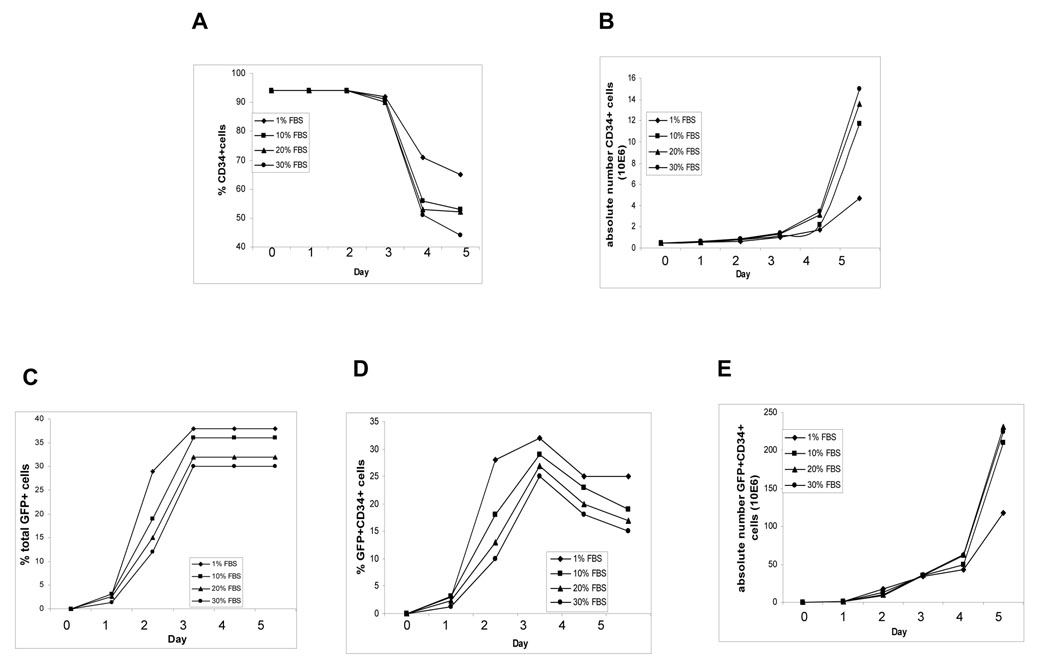
Fig. 1. Transduction of CB CD34+ cells with LVs.
Freshly isolated CB CD34+ cells were transduced with LVs for 5 hr. After extensive washing with PBS, the cells were cultured in IMDM containing SFT (100 ng/ml SCF, 100 ng/ml FL and 50 ng/ml Tpo) and FBS at 1, 10, 20, or 30%. Cells were counted and monitored daily for GFP expression for transduction efficiency daily. Cells were also stained daily with anti-CD34 antibody, and FACS was used to determine the percentage and numbers of CD34+ cells. A) % CD34+ cells, B) absolute number of CD34 + cells, C) % GFP+ cells, D) % GFP+CD34+ cells, and E) absolute number of GFP+CD34+ cells.

