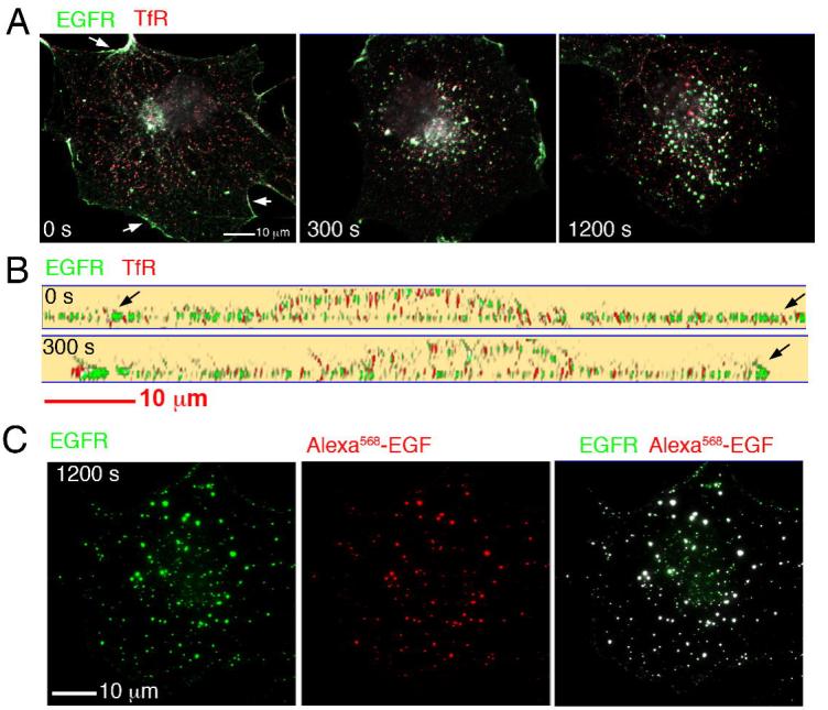
Figure 4.

A. COS-7 cells were exposed to unlabed EGF and Tf for the times indicated, fixed and stained with antibodies to the EGFR and the TFR. Optical sections were obtained at 250 nm intervals through the entire volume of the cell, and projected into a single 2D image. A clear demarcation of the cell edge was seen with antibodies to EGFR (arrows), but not to the TfR. B. 130 nm optical slices through the thickest part of the nucleus of cells treated and stained as in A. C. Cells were exposed to Alexa568-EGF for 20 min, fixed, and stained with antibodies to the EGFR. Optical sections were obtained at 250 nm intervals through the entire volume of the cell, and projected into a single 2D image.
