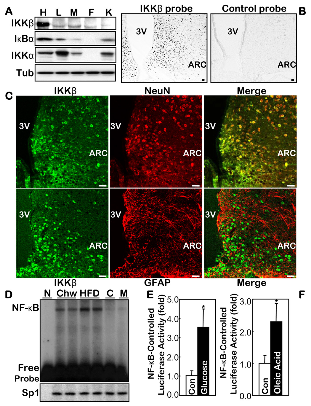
Fig. 1.
IKKβ/NF-κB in the hypothalamus and its relationship with overnutrition. A. Protein levels of IKKβ, IKKα and IκBα in the hypothalamus (H) vs. the peripheral organs including liver (L), skeletal muscle (M), fat (F), and kidney (K) from normal chow-fed C57BL/6 mice. Tub: tubulin. B & C. Distribution of IKKβ mRNA and protein in the hypothalamus was respectively profiled by probing the brain sections with anti-sense IKKβ oligonucleotides (B) and by co-immunostaining of IKKβ with NeuN or GFAP (C). The labeled sense sequence of IKKβ oligonucleotides was used as the control probe (B, right). Bar = 50 µm. D. DNA binding activity of NF-κB oligonucleotide and an irrelevant control (Sp1) was measured using EMSA in the hypothalamus from HFD- vs. chow-fed C57BL/6 mice. Cold competition (C) with 200-fold excess of unlabelled NF-κB oligo and mutant NF-κB probe (M) were used to assess the specificity of the NF-κB EMSA blots. N: non-specific protein (BSA). E & F. NF-κB-induced luciferase activity was measured in the hypothalamus harvested from the NF-κB reporter mice that received intra-third ventricle infusion of 1 mg glucose (E) or 40 mg oleic acid (F) over 6 hours. The controls (Con) represent the infusion of the empty vehicle or the same concentrations of sorbitol. (n = 4–5 per group, *p<0.05). 3V: third ventricle.

