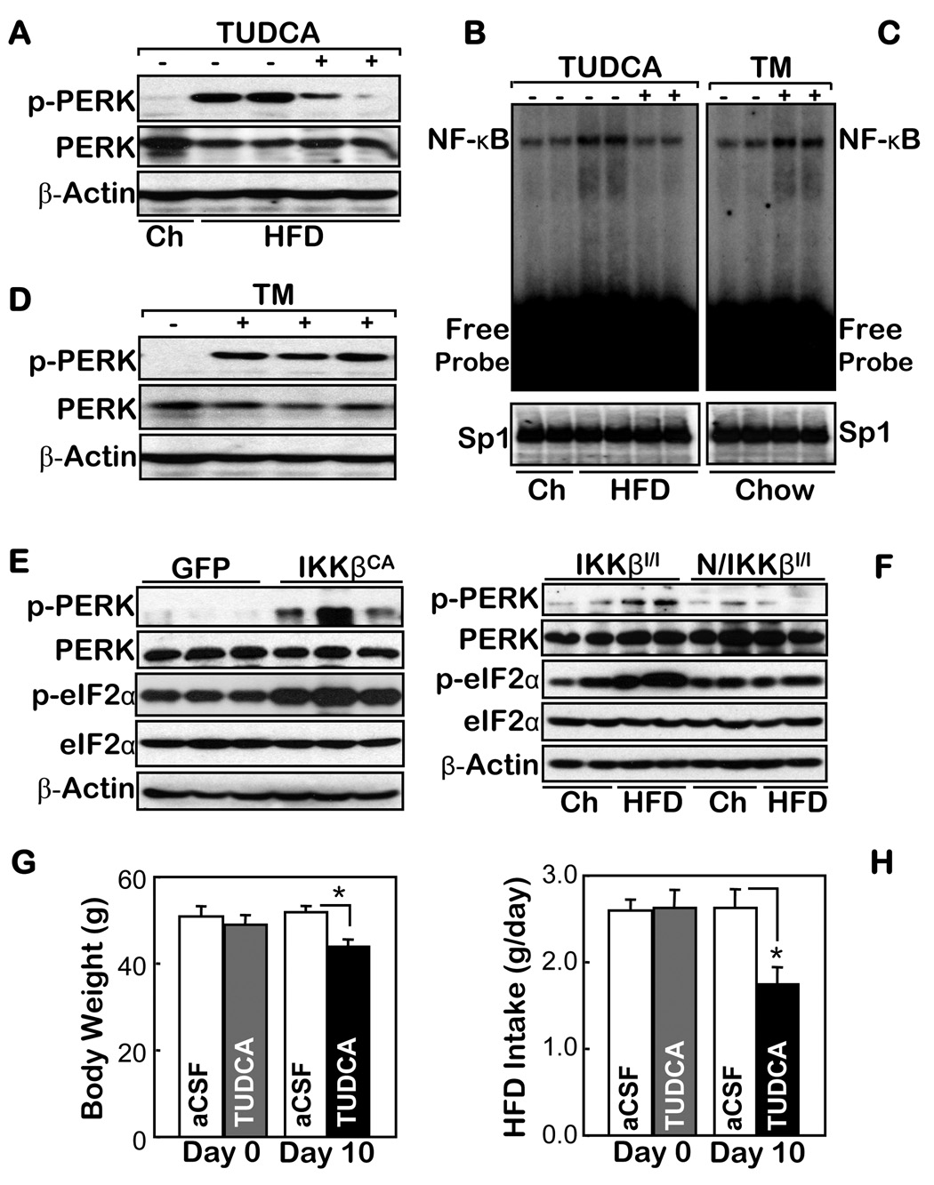
Figure 3.
The association of ER stress with IKKβ/NF-κB in the hypothalamus. A–D. Normal chow (Ch)-fed mice (A–D) and HFD-fed matched mice (A & B) received intra-third ventricle infusions of (+) 25 µg TUDCA (B) or 3 µg TM (C) vs. the vehicle control (−) over 2 hours (A–D). The markers of ER stress (A & D) and the DNA binding activities of NF-κB and an irrelevant control (Sp1) (B & C) in the hypothalamus of these mice were measured using Western blot and EMSA, respectively. E & F. The ER stress markers were measured in the hypothalamus of normal chow-fed mice that received intra-MBH injections of IKKβCA- vs. GFP-lentivirus (E) and normal chow- vs. HFD-fed Nestin/IKKβlox/lox mice (N/IKKβl/l) and their controls (IKKβlox/lox mice, IKKβl/l) (F). G & H. HFD-fed mice received daily intra-third ventricle injections of TUDCA ((5 µg/d) or the empty vehicle (aCSF) for 10 days. The body weight (G) and average food intake (H) of these mice were measured before (Day 0) and upon the completion of the 10-day therapy. (n = 5 per group; *p<0.05).

