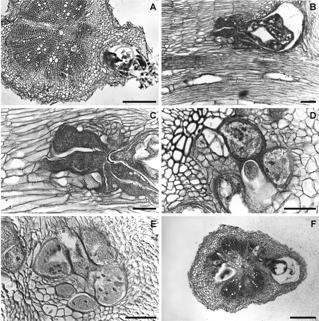
Fig. 7.
Infection and feeding site structures of Meloidogyne dunensis n. sp. in naturally infected sea rocket roots. A, F) Cross-sections showing nematode infections. B) Longitudinal section. C-E) details of nematode feeding-sites showing multinucleate giant cells with hypertrophied nuclei. Scale bars: A, F = 200 μm; B-E = 100 μm.

