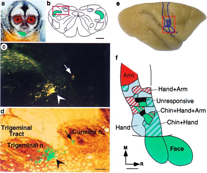
Figure 1.
The growth of terminals from trigeminal nucleus into the cuneate nucleus in an adult owl monkey 18 mo after partial transection of the dorsal columns. (a) The green overlay on the face of an owl monkey shows the region of the skin that received 29 5-μl injections of the transganglionic tracer B-HRP. (b) An outline of a coronal section of the lower brainstem showing the locations of cuneate nucleus (blue) and trigeminal nucleus (green). In normal monkeys, all of the tracer from skin injections such as those shown in a is confined to the expected locations in the trigeminal nucleus. The part of the brainstem that is shown at a higher magnification in c and d is boxed. (c) A dark-field photomicrograph of a brainstem section showing presence of the B-HRP label in the cuneate nucleus (arrow) after injections of the tracer in the skin of the chin. This label indicates the growth of trigeminal nucleus terminations into the cuneate nucleus. Normal terminations of the inputs from the face in the trigeminal nucleus are more numerous, and they are more strongly labeled (arrowhead). The labeled fibers in the upper left corner are in the trigeminal tract. (d) A section of the lower brainstem adjacent to that shown in c and stained for cytochrome oxidase activity. The cuneate and trigeminal nuclei and the trigeminal tract are marked, and the locations of the labeled terminals are marked in green. Images of the sections in c and d were digitized and overlaid in photoshop (Adobe Systems, Mountain View, CA) based on surface features and shared blood vessels. The label in c was converted to a channel mask on a separate layer and filled with green color, and the layer containing the section shown in c was deleted. (e) A dorsolateral view of an owl monkey brain showing location of the somatosensory area 3b. The hand and the face regions shown in f are boxed. (f) Reorganized somatotopy in area 3b as a result of the incomplete lesion of the dorsal columns of the spinal cord at C4/C5 level on the right side. In the deafferented hand region, there were responses to the stimulation of the chin in addition to the stimulation of the remaining inputs from the hand and arm. There were regions that responded to the stimulation of only chin (green), chin and hand (green and blue hatch), and the stimulation of the chin, hand, and arm (green, blue and red hatch). Other parts of the hand cortex responded to the stimulation of the hand (blue) or both hand and arm (blue and red hatch). There were small regions that did not respond to the stimulation of any part of the body (gray). The responses in the lateral part of area 3b that normally represents the face remained unaltered. R, rostral; M, medial; n., nucleus. (Scale bar, 1 mm in b and f, 200 μm in d.)

