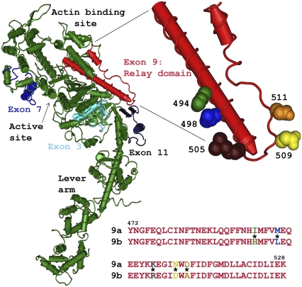
FIGURE 1.
Location and two alternative sequences of the Drosophila relay domain region. The relay domain (exon 9, red) and the three other variable regions (3, 7, and 11) encoded by Drosophila alternative exons (shades of blue) are mapped onto the chicken myosin S1 structure (green). The relay domain spans from the end of switch II, down toward the converter (dark blue), and back up toward the actin-binding site. The magnified region shows the position of the five amino acids in the relay that differ between the EMB and IFI myosin isoforms. The five amino acids shown are those of chicken skeletal muscle myosin, as the structure of Drosophila myosin has not been determined. The IFI (9a) and EMB (9b) alternative amino acid sequences are shown below the molecular structure. The color of the amino acid single letter corresponds to the space-filling amino acid in the relay magnified region; * signifies residues that are different between 9a and 9b.

