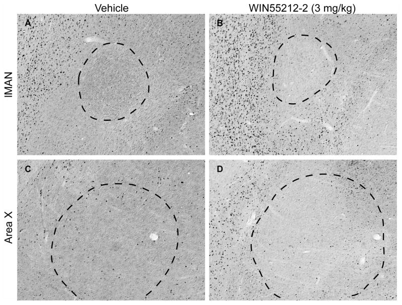
Figure 2.
Immunohistochemical staining of lMAN and Area X regions of rostral telencephalon with anti-c-Fos antibody as a function of vehicle (A and C) or WIN55212-2 (3 mg/kg, B and D) treatment. Medial parasaggital sections represent planes about 1.5 mm lateral from the midline. Rostral is left, dorsal is top, magnification is 100 X. Dark puncta represent stained nuclei. Note relatively low-level expression in lMAN (indicated by dashed outline in panels A and B) and Area X (outlined in panels C and D) relative to that within the caudal song regions HVC and RA (shown in Fig 3).

