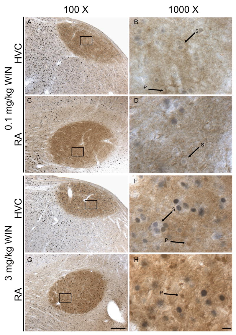
Figure 6.
Double-immunohistochemcial labeling with c-Fos and CB1 cannabinoid receptor antibodies. c-Fos-labeled nuclei are stained blue-grey, CB1 receptor staining is rust-brown. The pattern of anti-CB1 staining within HVC and RA consists of diffuse neuropil staining with distinct small and irregularly shaped puncta (indicated with arrows labeled ‘P’). c-Fos-labeled nuclei surrounded by unstained cytoplasm are indicated with arrows labeled ‘S’). Dorsal is top, rostral left. 100 X bars = 200 microns, 1000 X bars = 10 microns.

