Figure 1.
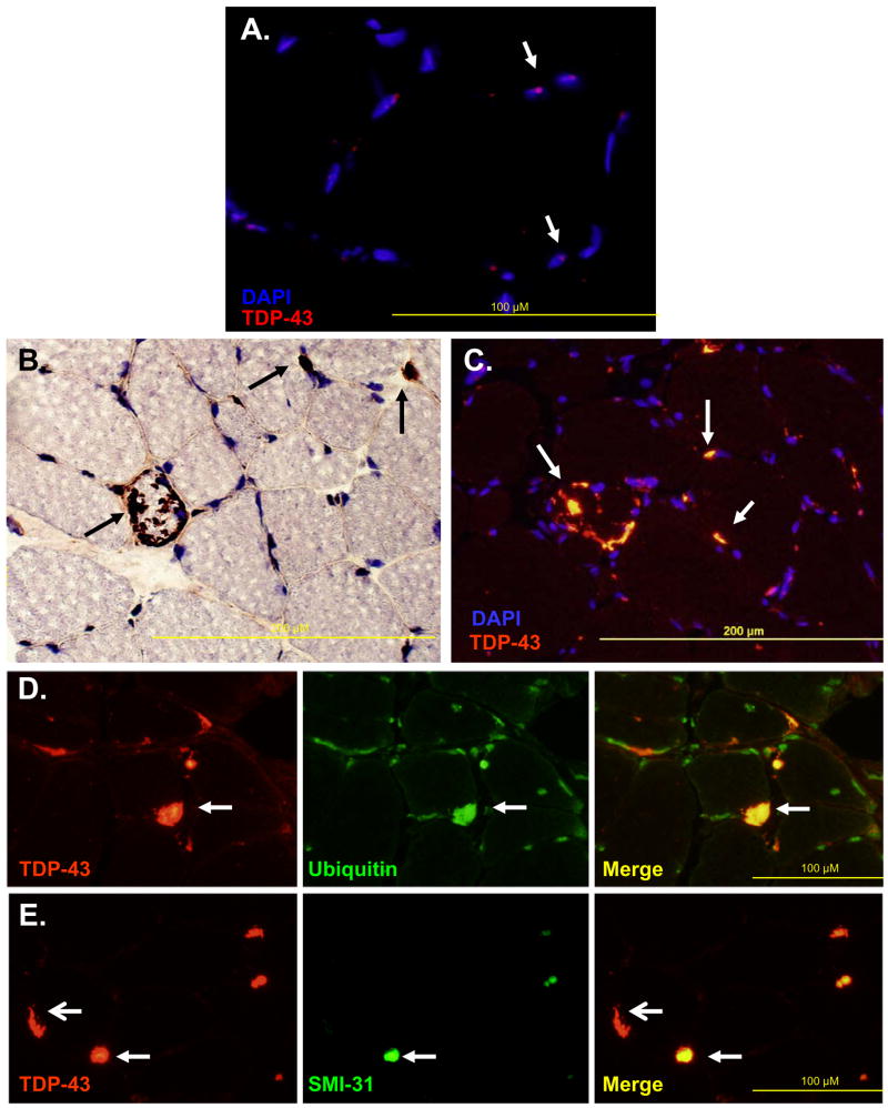
A) Normal muscle immunostained with anti-TDP-43 antibody and counterstained with DAPI to allow visualization of nuclei. Figure is overlay of anti-TDP-43 and DAPI images. Arrows denote blue nuclei with TDP-43 (red dots) in scattered myonuclei. B) IBMPFD patient tissue immunostained with anti-TDP-43 (brown) and counterstained with Congo red to allow visualization of nuclei and myofibers. Arrows denote large inclusions, some of which are peripherally based. C) Overlay of anti-TDP-43 (orange) and DAPI (blue) of IBMPFD patient tissue. Note that large peripheral inclusions do not localize within nuclei (arrows). D) IBMPFD patient tissue co-immunostained with anti-TDP-43 (red) and FK2 (green). Note that TDP-43 inclusions co-localize with FK2 (ubiquitinated proteins) (arrows). E) IBMPFD patient tissue co-immunostained with anti-TDP-43 (red) and SMI-31, an antibody against phosphorylated tau epitopes (green). Note that some TDP-43 inclusions co-localize with SMI31 (closed arrows) and others do not (open arrows).

