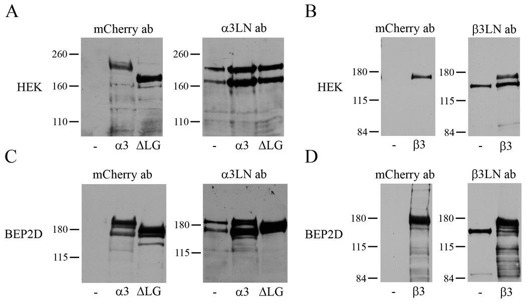
Fig. 2.
Extracts of HEKs (A,B) or BEP2D cells (C,D) were processed for immunoblotting. In A and C immunoblots of extracts of uninfected HEKs or BEP2D cells (-) or cells infected with virus encoding full-length, tagged α3 laminin (α3) or tagged α3ΔLG4-5 laminin (ΔLG) were probed with mCherry or α3 laminin subunit antibodies. In B and D immunoblots of uninfected HEKs and BEP2D cells (-) or cells infected with virus encoding tagged β3 laminin (β3) were probed with mCherry or β3 laminin subunit antibodies. Since there is differential expression of each of the tagged proteins in extracts of HEKs and BEP2D cells, the amount of protein loaded into the lanes of each gel in A and C was adjusted to optimize the reactivity with the mCherry probe. Molecular weight standards are marked at the left.

