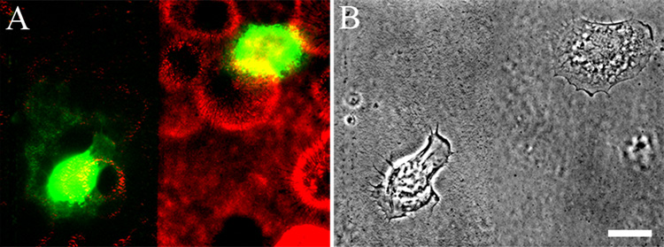
Fig. 6.
HEKs expressing mCherry-tagged β3 laminin were plated onto glass coverslips. 24 h later the confluent monolayer of cells was removed, leaving the matrix deposited by the cells behind (Gospodarowicz, 1984). Part of this matrix was scraped off as indicated. Subsequently, HEKs expressing YFP-tagged β3 laminin were plated sparsely onto the coverslips and the cells visualized 8 h later. In A the dual color image of the cells and their matrix is shown while in B, the phase contrast image is presented. The focal plane is close to the substratum-attached surface of the cells. Note that the cell on the left of the image in A has adhered to the region of the coverslip from which matrix was scraped off. This cell has elaborated a YFP-matrix. In contrast, the cell on the right of the image, which has adhered to the mCherry tagged matrix, has not assembled a YFP matrix, despite expressing YFP-tagged protein in the cytoplasm. This is representative of three different trials. Bar in B, 20µm .

