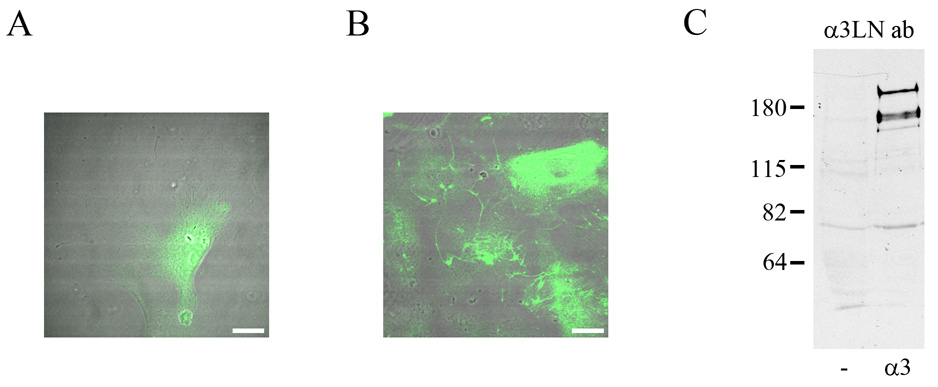
Fig. 7.
Uninfected ATII cells (A) of ATII cells infected with virus encoding expressing human full-length, tagged α3 laminin (B) were prepared for indirect immunofluorescence microscopy using an antibody specific for the human α3 laminin subunit. The focal plane is close to the substratum-attached surface of the cells. The fluorescent images have been overlaid on phase contrast views of the fixed and stained cells. The diffuse staining in the cells in A represents background. While there is no fibrous staining in A, the human α3 laminin subunit antibody stains a network of fibers in B. In C, extracts of uninfected ATII cells and ATII cells infected with virus encoding expressing human full-length, tagged α3 laminin were processed for immunoblotting using the human α3 laminin subunit. Equal amounts of protein were loaded in the two lanes. Molecular weight standards are marked at the left. Bars in A and B, 20µm .

