


Figure 1. NDGA inhibits survival of SKBR3 parental and trastuzumab-refractory breast cancer cells.

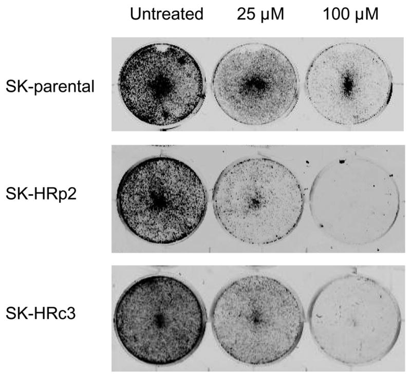

(A) Parental SKBR3 HER2-overexpressing breast cancer cells (SK-parental), SKBR3-derived trastuzumab-refractory pool 2 (SK-HRp2) and SKBR3-derived trastuzumab-refractory clone 3 (SK-HRc3) cells were treated with 0, 25, 50, or 100 μM NDGA for 72 h, at which point trypan blue exclusion analysis was performed. Cell viability is expressed as a percentage of untreated cells per line, with error bars representing standard deviation between replicates. Experiments were done in triplicate, and repeated at least twice. (B) SK-parental, SK-HRp2, and SK-HRc3 were treated with 0, 25, or 100 μM NDGA for 48 h. Drug-containing media was then removed, and cells were maintained in drug-free media for an additional week, at which point colonies were stained with methylene blue. Assays were repeated at least twice, in duplicate cultures each time. NDGA suppressed growth and survival of parental and trastuzumab-refractory HER2-overexpressing SKBR3 breast cancer cells. (C) SK-HRp2 and SK-HRc3 cells were treated with 0 or 100 μM NDGA for 48 h. Cells were lysed for protein and analyzed by ELISA for cytoplasmic histone-associated DNA fragments (mononucleosomes and oligonucleosomes) representative of apoptosis. Fold change in absorbance which reflects DNA fragmentation is shown relative to untreated cells, with error bars representing standard deviation between replicates. NDGA induced a 2-fold increase in DNA fragmentation, consistent with induction of apoptosis.