
Figure 4. NDGA inhibits IGF-I and HER2 signaling in trastuzumab-refractory cells.

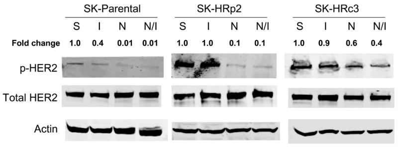


SK-parental, SK-HRp2, and SK-HRc3 cells were serum starved overnight. Cells were treated with NDGA (50 μM) for 1 h, followed by stimulation with IGF-I (100 ng/mL) for 20 min. S, serum-starved control; I, IGF-I stimulation alone; N, NDGA treatment alone; N/I, NDGA treatment followed by IGF-I stimulation. Total protein lysates were immunoblotted (50 μg) for (A) phosphorylated IGF-IR (p-IGF-IR) and total IGF-IR, (B) p-HER2 and total HER2, (C) p-Akt and total Akt, p-ERK1/2 and total ERK1/2. Actin served as a loading control. Blots were repeated at least twice to ensure reproducible results, and representative blots are shown. Bands were quantitated using NIH ImageJ software. NDGA blocked phosphorylation of IGF-IR and HER2 in parental and resistant cells. Akt phosphorylation was inhibited by NDGA in resistant cells, while ERK1/2 phosphorylation was reduced partially in SK-HRc3 cells. (D) SK-parental, SK-HRp2, and SK-HRc3 cells were untreated (0), treated with IGF-I (100 ng/mL) (I), NDGA (50 μM) (N), or combination NDGA (50 μM) plus IGF-I (100 ng/mL) (N+I) for 72 h prior to trypan blue exclusion analysis. Cell viability is expressed as a percentage of untreated cells per line, with error bars representing standard deviation between replicates. Trypan blue experiments were repeated at least twice, in triplicate cultures per group. NDGA suppressed cell survival in the presence of IGF-I in parental and trastuzumab-refractory cells. IGF-I stimulatory effects were more pronounced in resistant cells; IGF-I-mediated proliferation was inhibited by NDGA in resistant cells.