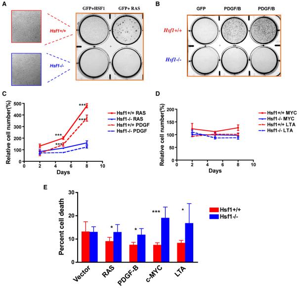
Figure 3. HSF1 Enables Cellular Transformation Initiated by Oncogenic RAS and PDGF-B.
(A) Hsf1-/- MEFs are relatively resistant to focus formation driven by oncogenic H-RASV12D. Immortalized MEFs were plated and transduced with retroviruses encoding the genes indicated. Foci were fixed and visualized by dye staining. The number of foci per well was quantified as shown in Figure S2. All experiments were repeated once with similar results.
(B) Hsf1-/- MEFs are relatively resistant to focus formation driven by the proto-oncogene PDGF-B.
(C) Hsf1-/- MEFs are refractory to proliferation driven by oncogenic RAS and PDGF-B. Equal numbers of immortalized Hsf1+/+ and Hsf1-/- MEFs were transduced with retroviruses encoding GFP, H-RASV12D,or PDGF-B. The cells were fixed on the indicated days and the number of cells per well determined by fluorescent DNA staining. Relative cell number was calculated by normalizing the values against the GFP-transduced group at each time point (mean ± SD, n = 5, ***p < 0.001, two-way ANOVA).
(D) Expression of c-MYC and LTA does not drive marked proliferation in immortalized Hsf1+/+ and Hsf1-/- MEFs.
(E) Hsf1-/- MEFs show no enhanced survival in response to RAS and PDGF/B expression but reduced survival in response to c-MYC and LTA expression. Viability of immortalized Hsf1+/+ and Hsf1-/- MEFs was determined by flow cytometry 36 hr after transduction. The data are presented as percent nonviable cells (mean ± SD, n = 5, *p < 0.05; ***p < 0.001, Student’s t test).

