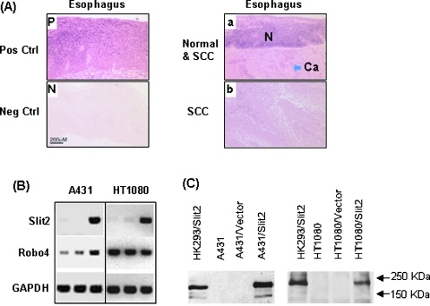
Figure 1.
Expression of Slit2 mRNA in normal and malignant esophageal tissues. (A) Slit2 mRNA was detected by in situ hybridization using digoxigenin-labeled antisense cRNA and positive cells are blue (Pos Ctrl). Sense cRNA was used as a negative control (Neg Ctrl). The normal esophageal epithelium (N) and SCC (Ca) are shown on the same section. Pictures were taken microscopically with a 10x objective. (B) Transcriptions of Slit2 and Robo4 in tumor cell lines HT1080 and A431 were detected by RT-PCR. The sample order of each cell line is parental, control vector-transfected, and Slit2-transfected cells. (C) Detection of cMyc-tagged Slit2 proteins. Slit2 proteins in culture supernatants of parental and transfected tumor cell lines were detected by Western blot using an anti-cMyc antibody. The supernatant of Slit2-transfected HK293 (HK293/Slit2) cells served as a positive control.

