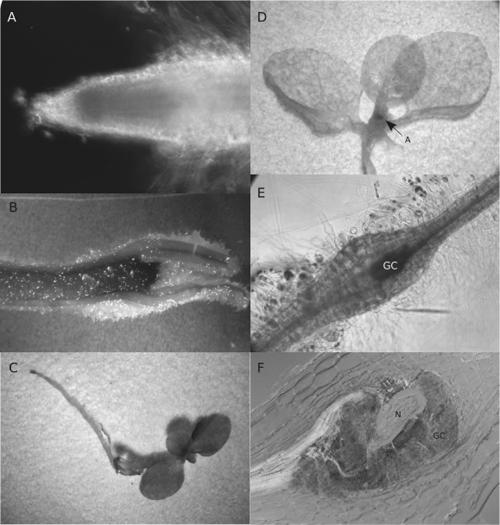
Fig. 2.
Histochemical staining for GUS activity in transgenic tobacco plants containing the Atcel1 promoter infected by the root-knot nematode, Meloidogyne incognita. A) Atcel1-driven GUS expression in a 7-day-old uninfected tobacco root. B) Atcel1-driven GUS expression in a shoot tip of a young uninfected tobacco seedling. C) Atcel1 promoter deletion construct B (harboring a 502 bp 5’ deletion, promoter fragment 1)-driven GUS expression in the shoot elongation zone of an uninfected tobacco plant. No activity is detectable in the roots. D) Whole-mount histochemical GUS assay of a Cel1-transgenic tobacco plant infected with M. incognita. Atcel1 activity is confined to the nematode feeding cells (not shown) and the plant elongation/ differentiation zones. A = shoot meristem (Construct B shown). E) Atcel1-driven GUS expression within M. incognita-induced giant cells four days post-inoculation of nematodes to an Atcel1-GUS transgenic tobacco root (construct A, Fig. 1). GUS activity is confined to the central region of the developing gall tissue. F) Sections (30 μm thick) through M. incognita-infected Atcel1-GUS tobacco roots after GUS staining. GUS expression is restricted to the giant cells induced by the nematode. N = nematode, GC = giant-cells,

