
Fig. 4.
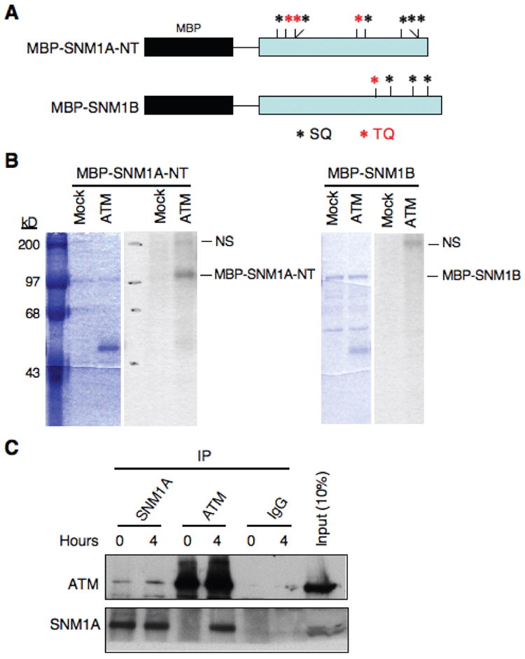
SNM1A is a phosphorylation substrate of ATM in vitro and interacts with ATM in vivo. (A) The SQ and TQ sites present in the N-terminus (NT) region of SNM1A and in full-length SNM1B are shown. (B) An IP kinase assay shows that recombinant MBP-SNM1A-NT (SNM1A residues 1-393), but not SNM1B is an ATM substrate. Left side of each panel shows Coomassie blue stained gels, and right sides show autoradiograms. “Mock” indicates an immunoprecipitation performed with a nonspecific IgG. “NS” indicates a nonspecific substrate of ATM. (C) Immunoblot showing reciprocal co-IP of ATM and SNM1A with or without IR treatment (10 Gy).