
Figure 3.
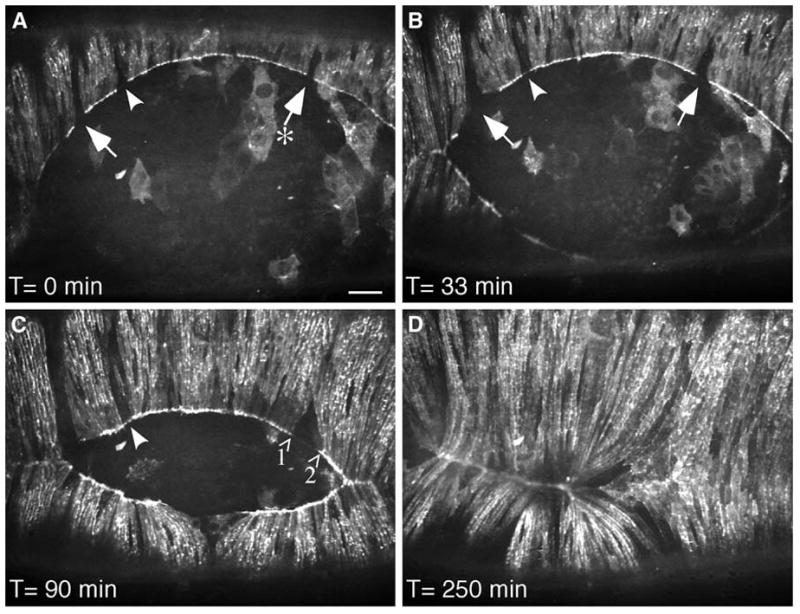
Tension created by myosin contraction is observed during dorsal closure of the epithelium in the fruit fly. In dorsal closure, two sheets of epithelial cells move towards each other and meet at the dorsal midline, leaving an eye-shaped opening. The epithelial sheets pull themselves together by three mechanisms: “zipping” inwards from the corners of the eye; constricting around the leading edge (like pulling on a drawstring); and using adhesion forces from the underlying cells. In flies with deletion of the homolog of vertebrate nonmuscle myosin heavy chain, dorsal closure fails and the embryos die. Transgenic overexpression of a GFP-myosin fusion protein in the myosin-null flies restores their ability to complete dorsal closure. However, in these flies, a few cells do not express the transgene and therefore do not express any myosin. On live fluorescent imaging of the flies during dorsal closure, these non-expressing cells are seen as gaps in the drawstring surrounding the opening between the epithelial cell sheets (A, arrows). As closure progresses, the space between GFP-myosin expressing cells clearly lengthens (A, arrow labeled with an asterisk; C, arrows labeled 1 and 2), visually demonstrating that contraction of myosin in the drawstring pulls on the neighboring cells to direct dorsal closure. Reproduced from Franke et al.56 with permission from Elsevier.