
Figure 3. Z-L3VS-induced formation of excessive daughter centrioles at maternal centrioles requires cyclin E, CDK2 and PLK4.
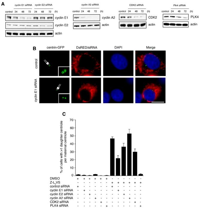
(A) Immunoblot analyses of U-2 OS/centrin-GFP cells transfected with either control siRNA duplexes (control) or siRNAs targeting cyclin E1, cyclin E2, cyclin A2, CDK2 or PLK4 for the indicated time intervals. Immunoblots for actin are shown to demonstrate loading of equal amounts of protein.
(B) Fluorescence microscopic analysis of U-2 OS/centrin-GFP cells transfected with control siRNA duplexes (top panels) or siRNAs targeting cyclin E1 (bottom panels) following treatment with 1 μM Z-L3VS for 48 h. Cells were co-transfected with DsRED fluorescent protein as transfection marker. Nuclei stained with DAPI. Scale bar indicates 10 μm.
(C) Quantification of U-2 OS/centrin-GFP cells transfected with the indicated siRNAs followed by either control treatment with 0.1% DMSO or 1 μM Z-L3VS for 48 h. Each bar represents mean and standard error of at least three independent experiments with a minimum of 100 cells counted per experiment.