
Figure 6. HPV-16 E7 rapidly stimulates the concurrent formation of more than one daughter centriole at maternal templates.
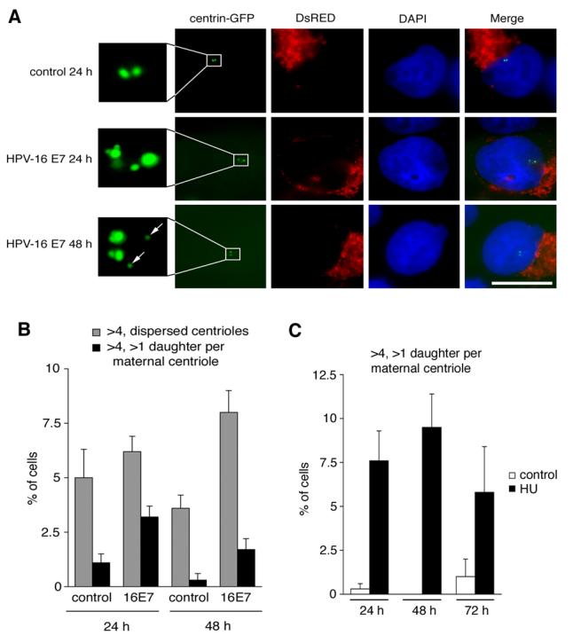
(A) Fluorescence microscopic analysis of U-2 OS/centrin-GFP cells transiently transfected with either empty vector (control) or HPV-16 E7 after 24 h or 48 h. Note the formation of two daughters at a single maternal centriole after 24 h in a HPV-16 E7-transfected cell (middle panels, insert). After 48 h, this pattern was less frequent and the majority of cells with abnormal centriole numbers showed a more dispersed arrangement (bottom panels, insert, arrows). Cells were co-transfected with DsRED fluorescent protein as transfection marker. Nuclei stained with DAPI. Scale bar indicates 10 μm.
(B) Quantification of U-2 OS/centrin-GFP cells with more than four centrioles in a random arrangement (gray bars) in comparison to cells with more than four centrioles and a concurrent formation of more than one daughter per maternal centriole (black bars). Cells were transfected with empty vector (control) or HPV-16 E7 for 24 h or 48 h. Each bar represents mean and standard error of at least three independent experiments with a minimum of 100 cells counted per experiment.
(C) Quantification of U-2 OS/centrin-GFP cells with more than four centrioles and concurrent formation of more than one daughter centriole at a maternal centriole after treatment with 1 mM HU for the indicated time intervals. dH2O-treated cells are shown as controls. Each bar represents mean and standard error of at least three independent experiments with a minimum of 100 cells counted per experiment.