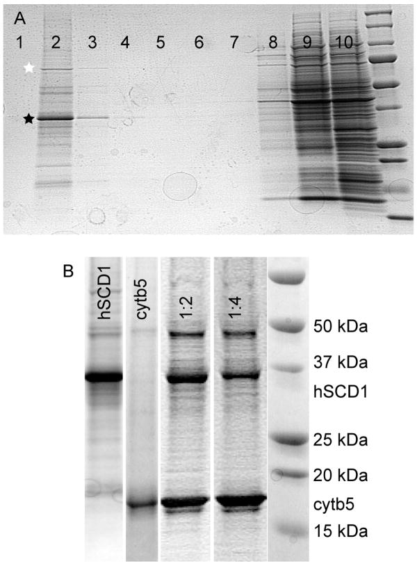
Fig. 3.
A, Coomassie-stained denaturing gel electrophoresis showing near complete incorporation of hSCD1 (black star) into synthetic liposomes from wheat germ cell-free translation. Proteoliposomes were loaded underneath a discontinuous Accudenz gradient and floated by ultracentrifugation. Bound protein, including hsp70 (white star) and hSCD1 floated (lanes 2 and 3), while unbound protein remained at the bottom (lanes 8–10). B, a comparison of proteoliposomes from floated fraction 2 containing hSCD1 (lane 1), cytb5 (lane 2), and a co-translation of hSCD1 and cytb5 (lane 3, 1 eq of hSCD1 mRNA with 2 eq of cytb5 mRNA; lane 4, 1 eq of hSCD1 mRNA with 4 eq of cytb5 mRNA).

