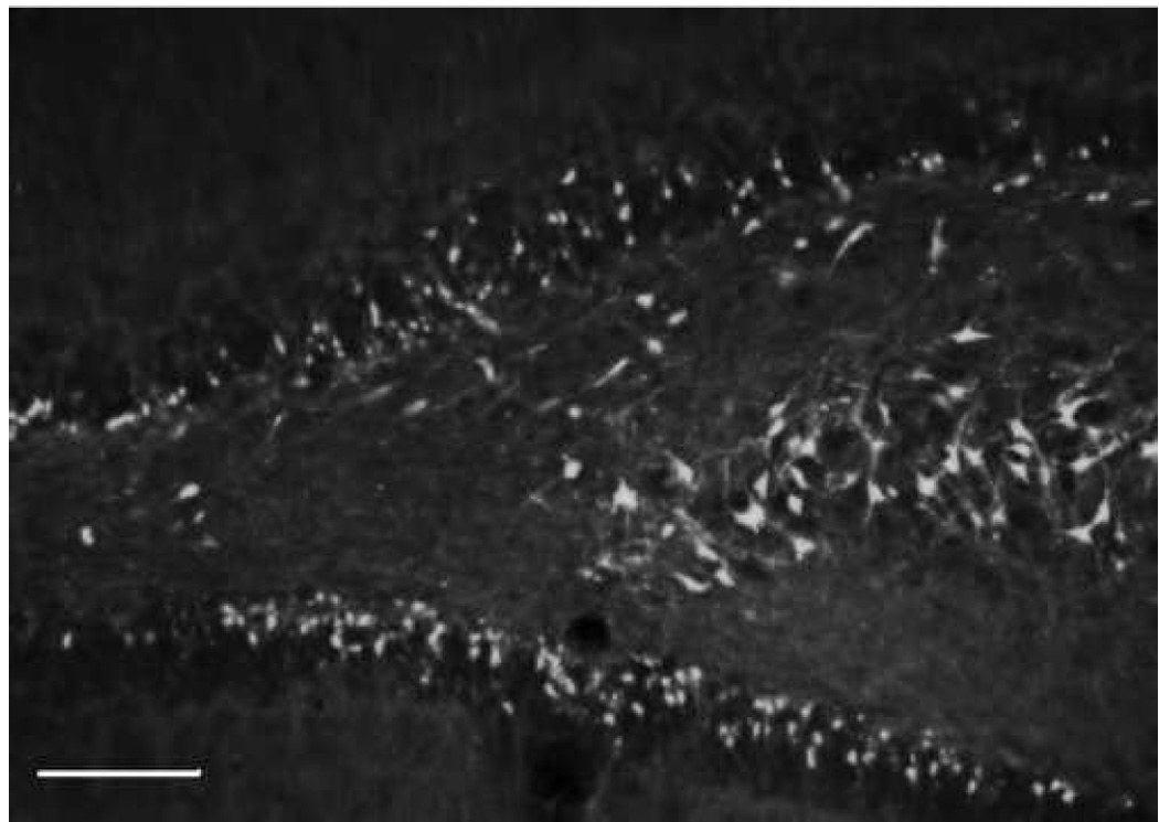
Fig. 9.

Ipsilateral coronal section through the hippocampal dentate gyrus and CA3 region at 24h following a unilateral cortical cortical contusion. Sections were stained with flour-Jade B (FJB) an anionic fluorescein derivative that specifically identifies degenerating neurons. Stereological techniques were used to determine the total number of FJB-positive neurons in different subregions of the hippocampal formation. Calibration bar = 500um
