Figure 4.
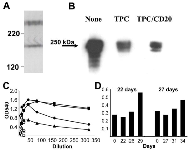
Characterization of high molecular weight non-Gal antigens detected by Group A antibody. A. High resolution 1D Western blot of high molecular weight antigens detected by IgG purified from a TPC treated recipient. B. Western blot analysis of antibody eluted from Group A rejected xenograft binding to purified pig fibronectin (1 μg/lane) electrophoresed in a 5% denaturing acrylamide gel. The treatment for each recipient is presented above the lane. C. ELISA analysis of pretransplant (open symbols) and necropsy serum (closed symbols) of Group A IgG binding to porcine fibronectin. No treatment (circle), TPC treated (square and triangle), TPC and Rituximab treated (diamond). D. ELISA analysis of anti-fibronectin IgG activity in 2% serum from Group B recipients that survived explant. The x-axis indicates the transplant day for each serum sample and the explant day is indicated above each graph.

