
Figure 5. Overexpression of IL-1β activates NF-κB in MDSCs in vivo.
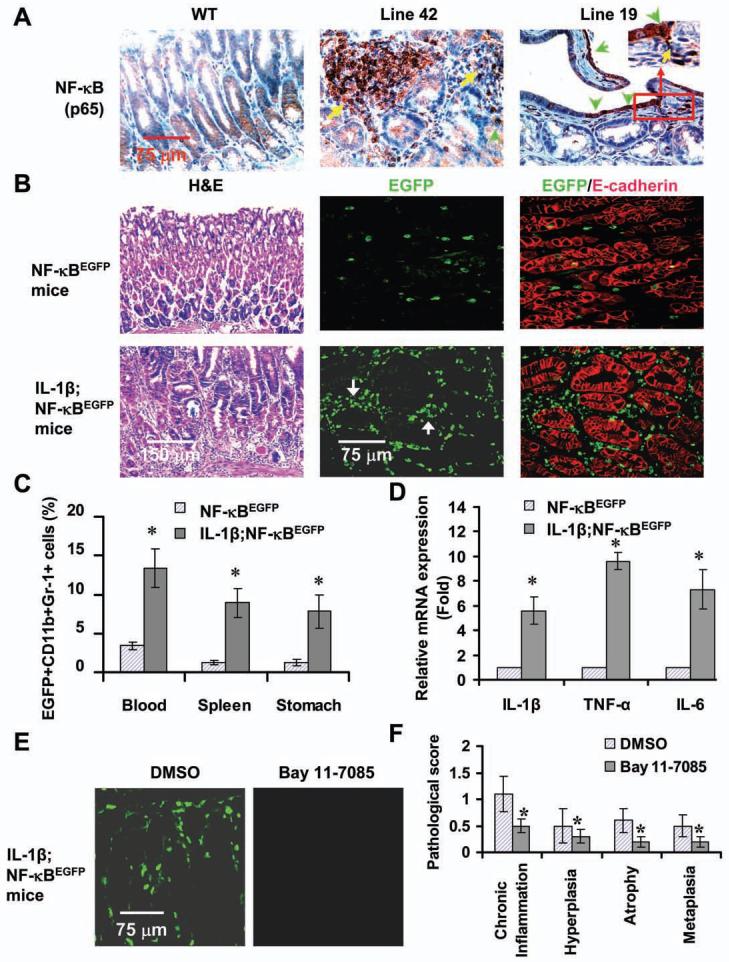
(A) NF-κB activation in the stomachs of IL-1β mice. Sections were stained a NF-κB p65 antibody. Arrows indicate p65+ cells. (B) Enhanced EGFP expression in IL-1β;NF-κBEGFP mice. Frozen gastric sections from 6 month old indicated mice were subjected to H&E staining and double staining with anti-EGFP (green) and E-cadherin (red) antibodies. The red staining indicates epithelial cells. Localization of EGFP+ cells is mostly confined to stromal region. (C) Increased frequencies of EGFP+ MDSCs in peripheral blood, spleen and stomach tissues in IL-1β;NF-κBEGFP mice as analyzed by FACS. The data represent the mean ± SD of 6 animals (*p < 0.05, vs NF-κBEGFP mice). (D) Increased expression of cytokines in stomach MDSCs in IL-1β;NF-κBEGFP mice. Stomach MDSCs sorted from 6 month old NF-κBEGFP and IL-1β;NF-κBEGFP mice were restimulated with PMA for 4 hours. mRNA expression was determined by real-time PCR. The data are normalized to MDSCs of NF-κBEGFP mice and represent the means ± SD of three independent experiments (*p < 0.01, vs NF-κBEGFP mice). (E) Blocking NF-κB activity by injecting i.p. Bay 11-7085 prevents EGFP expression in 3 month old IL-1β;NF-κBEGFP mice. Representative photos were taken from frozen gastric sections under fluorescence microscope. (F) Blocking NF-κB activity inhibits the development of gastritis. The pathological scores were graded in Bay 11-7085 or DMSO-treated IL-1β;NF-κBEGFP mice (*p < 0.05, vs DMSO treated mice, n = 8).