
Fig. 2. The Roc domain/tubulin interaction is guanine nucleotide independent.
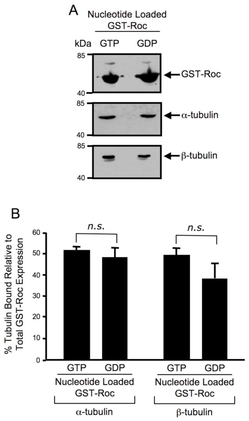
(A) Western blot analysis of guanine nucleotide loaded GST-Roc pull-down assay. GST-Roc recombinant protein was pre-loaded with either GTP or GDP prior to a pull-down assay with SH-SY5Y cell lysate. Precipitating proteins were resolved by 12.5% Tris-glycine SDS-PAGE, electrophoretically transferred to PVDF and Western blotted with GST, α-tubulin and β-tubulin antibodies. The Western blots shown are representative of three independent experiments. (B) Quantification of Western blot analysis of guanine nucleotide loaded GST-Roc pull-down assay. GST, α-tubulin and β-tubulin Western blots were quantified by densitometry and the percentage of α/β-tubulin bound was normalized to GST-Roc protein levels. Error bars represent standard error of the mean (SEM) for three independent experiments. n.s. is non-significant as assessed by a two-tailed unpaired Student’s t-test.