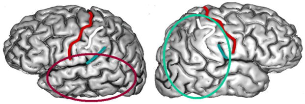
Figure 1.
Information processing maps in the temporal lobes (red oval) are differentiated from perisylvian auditory maps that are biased toward fast automatic digital processing in the time (temporal) domain, while parietal information processing maps (turquoise oval) differentiate from somatosensory and visual maps that are biased towards slower spatial reconstruction. Because of the anatomical asymmetry of the Sylvian fissure, temporal maps dominate left hemisphere processing and parietal maps dominate right hemisphere processing. Central sulcus, outlined in red, separates frontal and parietal lobes.

