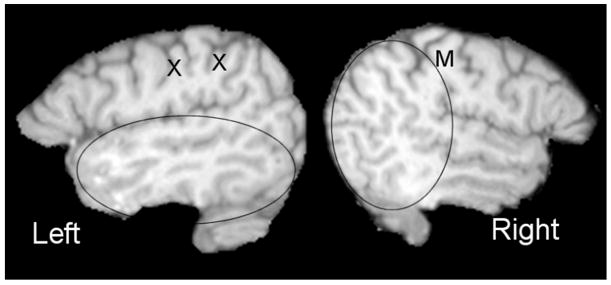
Figure 3.
Sagittal MRI images of the left and right hemispheres of a severely dyslexic man illustrating anomalous Sylvian fissure variants in both the left and right hemisphere. The X’s depict anomalous extra gyri interspersed between the central sulcus and the parietal planum. On the right, the planum parietale terminating the Sylvian fissure rises directly posterior to the central sulcus rather than the postcentral sulcus (compare the number of sulci descending from the superior surface anterior to the planum parietale on the left and right). These anomalies increase the asymmetries between the temporal and parietale lobes in the two hemispheres, leading, it is argued, to inefficient bottom up processing.

