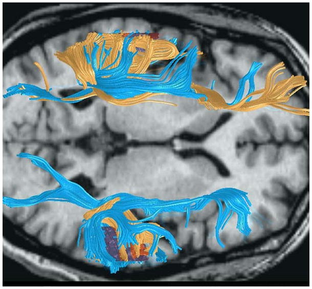
Figure 8.
This horizontal MRI image shows the overlain fiber tracings from diffusion tensor imaging of two men with different structural anatomy. The fibers depicted in yellow come from a man with normal leftward planar asymmetry. The fibers traced in blue come from a man with symmetrical plana temporale due to a large planum temporale in the right hemisphere (bottom of figure). Note the absence of connections between the temporal and frontal lobes in the right hemisphere in the man with normal asymmetry. We hypothesize that the parietal lobe dominates frontal circuits on the right when temporal lobe connections are missing. Image modified from (Leonard, Eckert, & Kuldau, 2006), used with permission of Blackwell Publishing.

