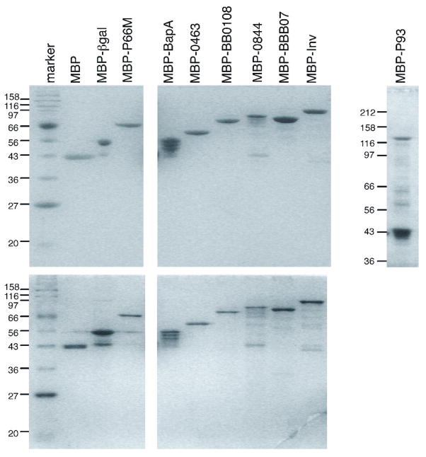
Figure 1. Electrophoretic analysis of recombinant MBP fusions to candidate integrin ligands.
Top panel: Samples of approximately 1.5 μg by BCA determination were stained with Coomassie brilliant blue. Bottom panel: Samples of approximately 0.3 μg stained with silver. Positions and sizes in kilodaltons of molecular weight markers are shown on the left. P93 was fractionated on a 10% polyacrylamide gel, the other proteins were fractionated on 12% gels.

