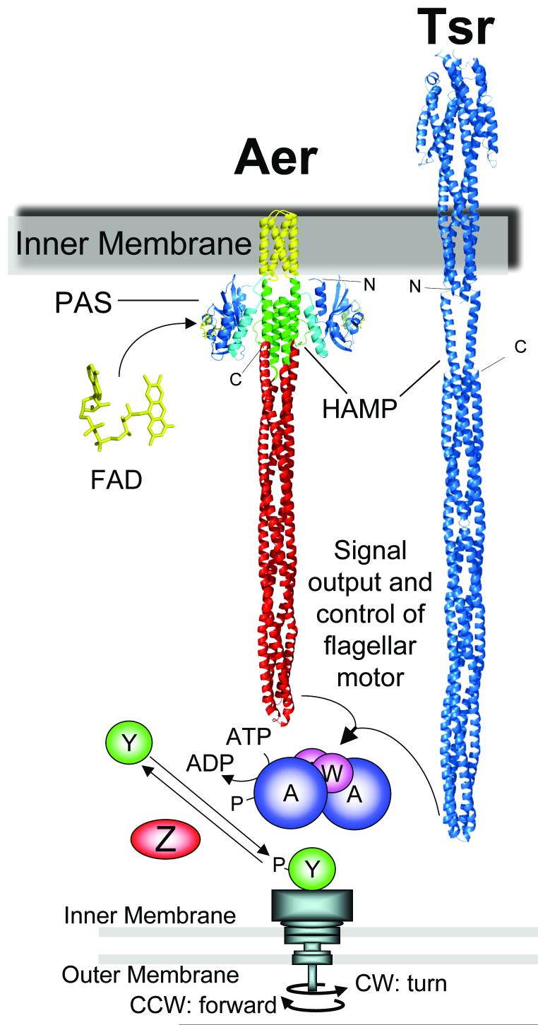
Fig. 1.
Comparison of the E. coli aerotaxis receptor, Aer, and the Tsr chemoreceptor, an alternative receptor for aerotaxis. The HAMP domain is shown in Tsr as an extended helix and in Aer as a four-helix bundle to illustrate different models for HAMP structure. Receptors shown are in silico constructions modeled from resolved structures and disulfide crosslinking studies: Aer-PAS [blue, (Key et al., 2007)]; F-1 (cyan); Aer-transmembrane [yellow, (Amin et al., 2006)]; Aer-HAMP [green, (Hulko et al., 2006)]; Aer-signalling [red, (Park et al., 2006)]. Tsr coordinates are from S-H. Kim as described in (Chi et al., 1997; Kim et al., 1999). The chemotaxis signalling pathway that controls flagellar rotation is also shown. Abbreviations: A, CheA protein; W, CheW; Y, CheY, Z, CheZ; P, phosphate; IM, inner membrane; OM, outer membrane.

