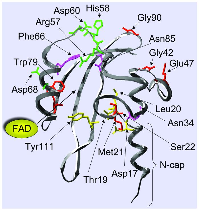
Fig. 3.
In silico model of the Aer PAS domain, highlighting residues involved in aerotaxis. The cleft in which FAD binds is shown. Replacement of the residues shown produced a null aerotaxis phenotype (red), a loss of FAD binding and a null phenotype (green), an inverted response (yellow), and a CW-signalling bias (magenta).

