
Fig. 6.
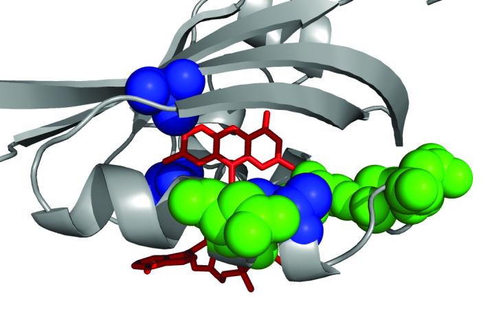
A view of the FAD-binding cavity in Aer PAS showing residues critical for FAD binding (green), and location of general suppressors (blue) of HAMP[AS-2] null mutants. FAD coordinates are modeled on the crystal structure of the NifL protein.
Official websites use .gov
A
.gov website belongs to an official
government organization in the United States.
Secure .gov websites use HTTPS
A lock (
) or https:// means you've safely
connected to the .gov website. Share sensitive
information only on official, secure websites.
A view of the FAD-binding cavity in Aer PAS showing residues critical for FAD binding (green), and location of general suppressors (blue) of HAMP[AS-2] null mutants. FAD coordinates are modeled on the crystal structure of the NifL protein.