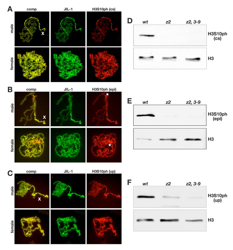
Fig. 2. Immunocytochemistry and immunoblot characterization of three different H3S10ph antibodies.

(A-C) Acid-free polytene chromosome squash preparations from male and female third instar larvae double labeled with antibodies to JIL-1 (in green) and H3S10ph (in red). H3S10ph labeling with antibody from Cell Signaling (cs) is shown in (A), from Epitomics (epi) in (B), and from Upstate (up) in (C). Composite images (comp) of the labelings are shown to the left. The labeling of all three H3S10ph antibodies shows co-localization with JIL-1 and upregulation on the male X chromosome (X). The Epitomics H3S10ph antibody in contrast to the two other antibodies showed strong labeling of the chromocenter (B, asterisks). The images in (A) are projection images from confocal sections. (D-F) Immunoblots of protein extracts from salivary glands from wild-type (wt), JIL-1z2/JIL-1z2 (z2), and JIL-1z2/JIL-1z2 Su(var)3-906 (z2, 3-9) larvae labeled with H3S10ph antibody from Cell Signaling (D), Epitomics (E), and Upstate (F). H3S10ph antibody labeling by all three antibodies is greatly reduced in JIL-1 null mutant backgrounds. Labeling with histone H3 (H3) antibody was used as a loading control.
