Figure 4.
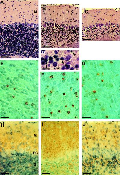
Neuropathological changes in the cerebella of Tg(PG14+/+) mice. Hematoxylin and eosin-stained sections showing the cerebellar cortex of mice of 22 days (A), 100 days (B and D), and 183 days (C) of age. M, molecular layer; PC, Purkinje cell layer; G, granule cell layer. Note the dramatic decrease in the number of granule cells with age. Arrowheads in D indicate pyknotic nuclei. ISEL-stained sections showing positively stained cells (brown) in the granule cell layer from mice of 31 days (E), 53 days (F), and 181 days (G) of age. PrP immunostaining of cerebellar cortex from mice of 22 days (H), 100 days (I), and 181 days (J) of age. Note the small punctate deposits of PrP. Scale bars are: 50 μm (A–C), 10 μm (D), 13 μm (E–G), 32 μm (H–J).

