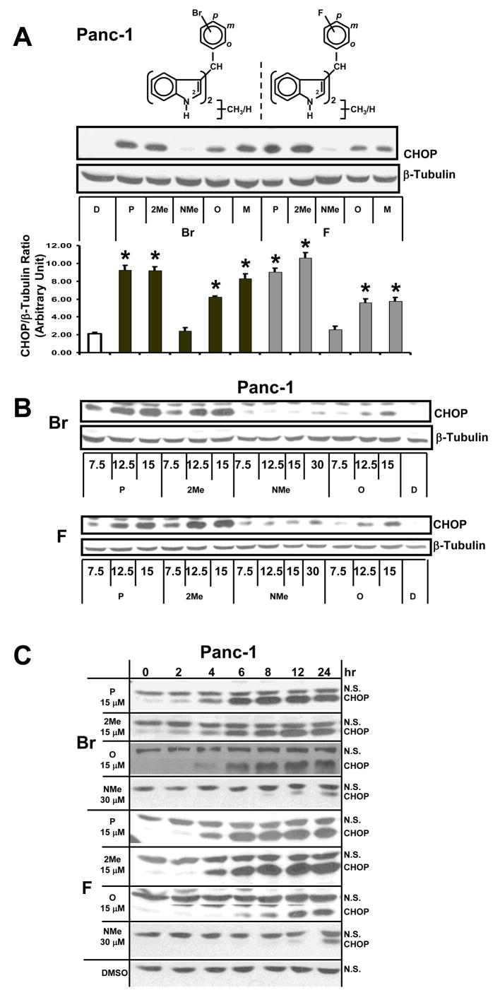
Figure 3. Structure-dependent activation of CHOP by DIM-C-pPhBr and DIM-C-pPhF analogs.
Structure-(A), dose-(B) and time-(C) dependent activation of CHOP in Panc-1 cancer cells. Cells were treated with either DMSO (D) or 15/30 μM concentrations of the C-DIM derivatives as indicated for 12 hr (A, B) or different periods of time (C), and changes in protein expression were determined by Western blot analysis of whole cell lysates as described in the Materials and Methods. Protein levels were measured with Image J and normalized to β-tubulin. All experiments were carried out at least 3 times and results in (A) are means ± SD for 3 replicate determinations and significant (p< 0.5) induction of CHOP is indicated by an asterisk.

