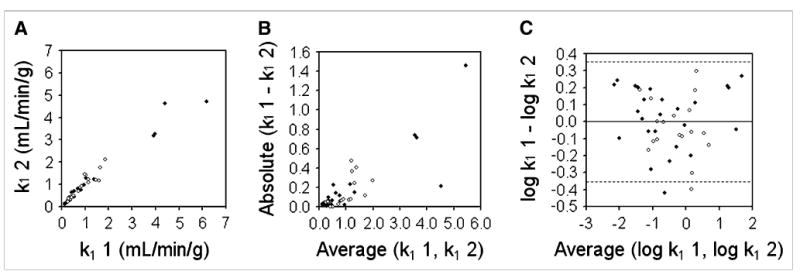
Figure 3.

Bland–Altman analysis of reproducibility of k1 for tumors (TBF). (A) Replicate measures of k1 are plotted against each other. Solid symbols represent data acquired on Advance scanner; open symbols represent data acquired on Discovery RX scanner. (B) Absolute difference between replicate k1 measurements are plotted as function of their mean and show clear proportionality. (C) After log transformation, dsd for k1 was calculated as 0.178. Dashed lines denote 95% CIs on either side of mean (1.96 × dsd).
