
Figure 4.
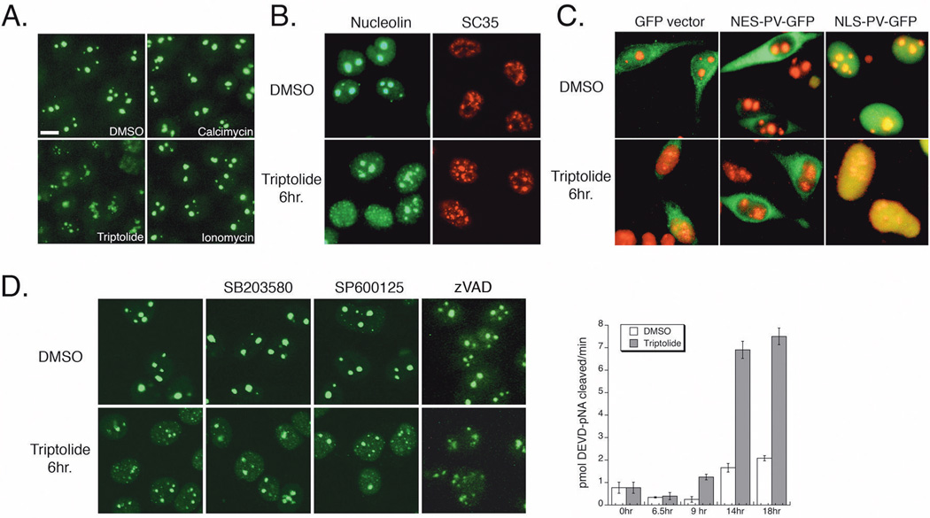
Nuclear Changes are Independent of Calcium, Stress Kinases, and Caspase Activation. A) HeLa cells were incubated for 6 hours with DMSO, 100 nM triptolide, 5 µM calcimycin, or 3 µM ionomycin before fixation and nucleolin detection by indirect immunofluorescence (25X magnification). Scale bar is 10 µm. B) HeLa cells were incubated in calcium free media for 16 hours before addition of DMSO or 100 nM triptolide for 6 hours. Cells were stained for SC35 (red) or nucleolin (green) by indirect immunofluorescence (confocal microscopy, 40X). C) HeLa cells were transiently transfected with GFP vector, nuclear excluded (NES) or nuclear localized (NLS) parvalbumin (PV) – GFP constructs for 24 hours before a 6-hour incubation with DMSO or 100 nM triptolide. GFP localization is in green, nucleolin is detected by indirect immunofluorescence (red). Images acquired separately under 40X magnification and artificially merged. D) HeLa cells were treated with DMSO, 5 µM SB203580, 10 µM SP600125, or 2 µM zVAD-fmk ± 100 nM triptolide for 6 hours. Nucleolar integrity was detected by indirect immunofluorescence of nucleolin (green). Graph represents caspase-3 activity in HeLa cells treated with DMSO or 100 nM triptolide over an 18-hour time course. Cell lysates were prepared and activity assessed by cleavage of the colorimetric substrate, DEVD-pNA. Data represent Mean±SE, n=3.