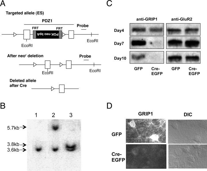
Figure 1.
Mutant mice harboring mutations in GRIP1 and GRIP2. A, Schematic representation of the GRIP1 conditional targeting strategy. The targeted allele of GRIP1 after homologous recombination is shown at the top, with two exons (white boxes) encoding the first PDZ domain. The neor cassette (black box; PGK-neo bpA), flanked by FRT sequences (gray boxes), was inserted between the two exons. Two loxP sites (arrowheads) were inserted before the first PDZ1 exon and immediately after the neor cassette; thus, expression of Cre recombinase would result in deletion of the first PDZ1 exon and cause a frameshift mutation that introduces an immediate stop codon in the GRIP1 transcript. The neor cassette was deleted by crossing the mutant mice with flippase transgenic mice. Two EcoRI restriction sites and the region for the probe for Southern blot analysis are also shown. B, Southern blot analysis of GRIP1 conditional knock-out mice. The genomic DNA was digested by EcoRI and detected with the probe illustrated in A. Wild-type mice gave a fragment of 3.6 kb, whereas the neor(+) mice extend the size of the fragment to 5.7 kb. When the neor cassette is deleted, the probed fragment is decreased to 3.8 kb. Lane 1: wt/wt, 3.6 kb; lane 2: heterozyogotes with the targeting vector, GRIP1(flox)neor (+)/wt, 5.7/3.6 kb; lane 3: heterozygotes after neor deletion, GRIP1(flox/+)neor (−)/wt, 3.8/3.6 kb. C, Cre lentivirus-mediated GRIP1 deletion in mouse cortical neurons. Cortical cultures were prepared from GRIP1(flox/flox) and GRIP2(−/−) mice and infected with EGFP or EGFP-IRES-Cre lentiviruses. Four, seven, and ten days after infection, neurons were harvested to detect GRIP1 expression level. Western blots of GRIP1 using anti-GRIP1 antibody (left) and GluR2 (right) are shown. D, GRIP1 immunocytochemistry after Cre-lentivirus infection. GRIP1(flox/flox) and GRIP2(−/−) cortical neurons were infected with EGFP-IRES-Cre or EGFP lentiviruses for 7 d and fixed and stained with anti-GRIP1 monoclonal antibody (left). Differential interference contrast (DIC) images of the same neurons are shown on the right.

