
Figure 6. Expression of native PPARδ is elevated in benign and malignant human thyroid tumors and correlates with cell proliferation.
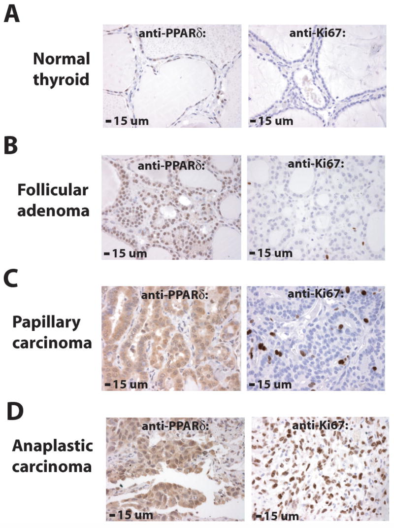
The expression of PPARδ was determined by immunohistochemistry on tissue microarrays that contained six different classes of human thyroid tumors. Normal thyroid tissues from the same paraffin blocks were used as controls. Levels of PPARδ and the proliferation marker Ki67 were quantified as described in Materials and Methods. (A) PPARδ was expressed at moderate levels in the nuclei and low levels in the cytoplasm (mean ACIS score 75.19) of normal thyroid cells. Ki67 was detected in 1.26% of nuclei in normal thyroid cells. (B) PPARδ was expressed at higher than normal levels (mean ACIS score 208.44, p<0.0001) in nuclei of benign thyroid follicular adenomas. Ki67 was detected in 2.02% of nuclei in follicular adenomas (p=0.0219). (C) PPARδ was expressed at higher than normal levels (mean ACIS score 394.11, p<0.0001) in the nuclei and cytoplasm of well-differentiated papillary thyroid carcinomas. Ki67 was detected in 4.53% of nuclei in papillary carcinomas (p<0.0001). (D) PPARδ was expressed at highest levels (mean ACIS score 438.60, p<0.0001) in poorly differentiated anaplastic thyroid carcinomas. Ki67 was detected in 31.18% of nuclei in anaplastic carcinomas (p<0.0001). PPARδ and Ki67 immunoreactivity is brown and the nuclear counterstain is blue (see also Table 1).