
Figure 6. p38α signaling limits the activation of other MAP kinase signaling pathways.
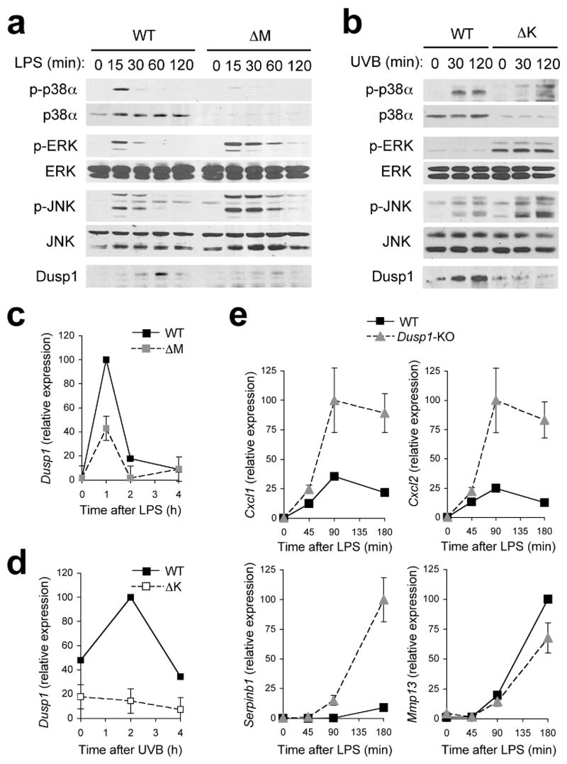
(a, b) Whole cell lysates from BMDMs treated with LPS (a), and keratinocytes irradiated with UVB (b) were prepared after the indicated durations of stimulation and analyzed by immunoblotting with antibodies against the proteins indicated on the left. Data are representative of at least five independent experiments.
(c, d) Dusp1 mRNA expression in BMDMs (c) and keratinocytes (d) at the indicated time points following LPS treatment (c) and UVB irradiation (d) was analyzed by qPCR. Data are representative of two independent experiments.
(e) Expression of genes in WT and Dusp1-KO BMDMs at the indicated time points following LPS treatment was analyzed by qPCR. Data are representative of two independent experiments.