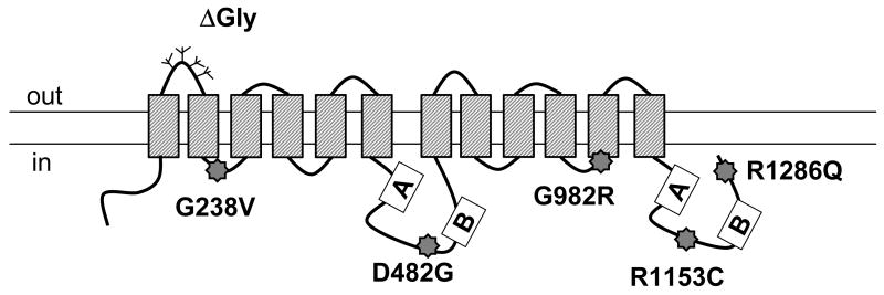
Fig.1. The locations of PFIC II mutations and ΔGly in Bsep.
The positions of G238V, D482G, G982R, R1153C and R1286Q are indicated by star signs in a predicted topology model of rat Bsep. The Walker A and B regions are shown in boxes and a number of potential glycosylation sites are indicated by the branch structures in the first extracellular loop of Bsep. ΔGly represents a mutation that lacks the potential glycosylation sites. The orientation of the plasma membrane is indicated.

